Often might not actually represent calcifications 6 most specific finding associated with. Irregular hypoechoic masses on breast ultrasound are usually considered suspicious lesions.
Benign And Malignant Characteristics Of Breast Lesions At Ultrasound Radiology Reference Article Radiopaedia Org
Three-dimensional scanners with the capability of reproducing high-resolution images in the coronal plane provide additional important information.

. Other histological aspects of galactocele breast lesions. In one report breast fibromatosis showed an isointense mass on unenhanced T1-weighted images heterogeneously low- to high-intensity mass on fat-suppressed T2-weighted images and. The image below shows an ill-defined border an irregular shape microlobulations and spiculations.
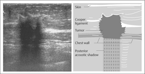
Oral cholecystography remains an excellent method of gallstone detection but its role has been limited due to the advantages of US. This could be due to an air collection from gallbladder rupture see labeled ultrasound below. Similarly the appearance of fibromatosis on ultrasound resembles malignancy appearing as an irregular hypoechoic mass with posterior acoustic shadowing.
Ultrasound evaluation of each renal unit is influenced by the orientation of the kidney in the retroperitoneum. The phenomenon of acoustic shadowing sometimes somewhat tautologically called posterior acoustic shadowing on an ultrasound image is characterized by a signal void behind structures that strongly absorb or reflect ultrasonic wavesIt is a form of imaging artifactThis happens most frequently with solid structures as sound conducts most rapidly in. The lower pole of the right kidney is 15 lateral to the upper pole in the coronal plane.
In the longitudinal view the standard orientation of the image should be with the superior pole of the testis to the left and the inferior pole to the right on the monitor screen Fig. This would all be highly predictive of invasive ductal carcinoma and the lesions would be need a biopsy for. Punctate echogenic foci without posterior shadowing.
If the lesions combine other features of malignancy such as spiculated margin nonparallel orientation and posterior shadowing they are considered moderate and highly suspicious for malignancy BI-RADS categories 4b and 4c or highly suggestive of malignancy. Posterior features represent the attenuation characteristics of a mass with respect to its acoustic transmission also of additional value. The characteristic US findings of gallstones are a highly reflective echo from the anterior surface of the gallstone mobility of the gallstone on repositioning the patient and marked posterior acoustic shadowing.
Figures 52 and 53 demonstrate the anatomic position of the right and left renal unit. Although calcification can be seen in both benign and malignant processes it is the ultrasound feature most closely associated with malignancy 1. The second image above focuses on this area and.
Alone it has little specificity. The lesion also appears to be taller-than-wide with an angular margin. Posterior acoustic enhancement is an indeterminate US finding that can be associated with a variety of entities including normal.
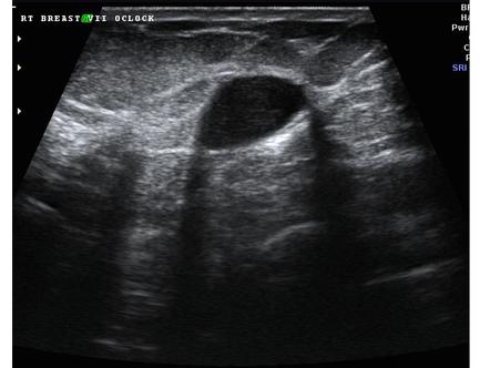
Posterior acoustic shadowing is a suspicious finding and may be seen in cases of invasive carcinoma postoperative scar complex sclerosing lesion or macrocalcifications and may even be seen in patients with dense breast tissue. Typically a malignant lesion presents as a hypoechoic nodular lesion which is taller than broader and has spiculated margins posterior acoustic shadowing and microcalcifications Figure Figure8A 8A F. The renal hilum is rotated approximately 30 posterior to the horizontal coronal plane when.
The American Institute of Ultrasound in Medicine AIUM guidelines recommend that scrotal ultrasound should be performed in at least two planes. To the right side of the image near the gallbladder fundus there is also shadowing but no evidence of stones. The first shows a large gallstone with posterior shadowing that is possibly impacted in the gallbladder neck.
Additionally there is often mild posterior shadowing distal acoustic enhancement. In other words the mammogram and the ultrasound might look confusing to the radiologist. Malignant masses can show posterior acoustic shadowing on ultrasound images.
The Relationship Between Lateral Acoustic Shadow Feature On Ultrasound Download Scientific Diagram
![]()
Transverse Ultrasound Of The Left Breast Demonstrates An Irregular Download Scientific Diagram

Ultrasound Image Of A Breast Cancer With Irregular Borders Angular Download Scientific Diagram
Basic Principles Radiology Key

Pdf Distinguishing Lesions From Posterior Acoustic Shadowing In Breast Ultrasound Via Non Linear Dimensionality Reduction Semantic Scholar

Mediconotebook Posterior Acoustic Shadowing And Enhancement

Posterior Acoustic Shadowing In Benign Breast Lesions Weinstein 2004 Journal Of Ultrasound In Medicine Wiley Online Library

Posterior Acoustic Shadowing In Benign Breast Lesions Weinstein 2004 Journal Of Ultrasound In Medicine Wiley Online Library
0 comments
Post a Comment